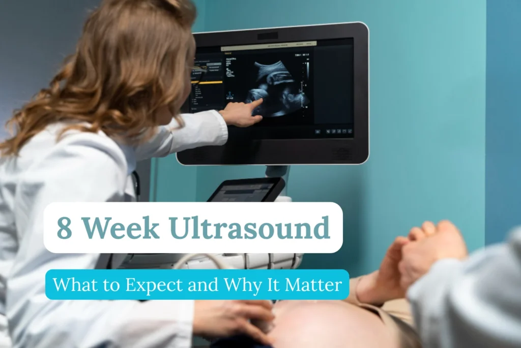
If you are 8 weeks pregnant, your doctor will likely recommend one of the first major milestones in prenatal care—the 8 week ultrasound. For many women, this appointment is filled with mixed emotions: excitement about finally seeing their baby on the screen, and a little anxiety about what the results may reveal. Both feelings are completely normal.
This early scan is more than just a first look at your little one. It plays a crucial role in confirming the pregnancy, checking your baby’s heartbeat, and making sure development is on track. In this guide, we’ll cover everything you need to know about the 8 weeks pregnant ultrasound—what to expect, what it looks like, why it matters, and the different types of ultrasounds you may have during pregnancy.
The ultrasound at 8 weeks is considered one of the most important first steps in prenatal care. International obstetrics guidelines recommend an early scan between the 7th and 9th week because it serves several key purposes: confirming that the pregnancy is inside the uterus, verifying the embryo’s heartbeat, and providing accurate dating of gestational age.
For expectant parents, this moment is both medical and deeply emotional. Medically, it provides reassurance about the health and progress of the pregnancy. Emotionally, it’s often the very first time parents see their baby’s heartbeat—a milestone that makes the journey feel real.
Medical Goals and Importance of First-Trimester Screening
The 8 weeks pregnant ultrasound is not only about getting a first glimpse of the baby. It has clear diagnostic goals that guide your healthcare provider:
- Vitality test:
- The detection of cardiac activity is the most important indicator of an intact pregnancy.
- A heartbeat at this stage significantly lowers the risk of miscarriage.
- Localization:
- Confirms that the pregnancy is intrauterine (within the uterus).
- Excludes an ectopic pregnancy, which can be life-threatening if untreated.
- Dating the pregnancy:
- By measuring the crown-rump length (CRL), doctors can determine the exact gestational age.
- This method is more reliable than using the date of the last menstrual period, especially in women with irregular cycles.
- Number of embryos:
- Determines whether you are carrying a singleton or multiples (twins, triplets, etc.).
- Early detection is crucial for monitoring high-risk pregnancies.
What Happens at 8 Weeks of Pregnancy?
At this stage, both your body and your baby undergo rapid development.
- Baby’s size: Around 1.4–2.0 cm (0.55–0.8 inches) long, comparable to a kidney bean.
- Heartbeat: Typically ranges from 120 to 180 beats per minute, visible as a flickering motion.
- Neural development: The neural tube (which later becomes the brain and spinal cord) is closing.
- Organ formation: The foundations of the heart, brain, liver, and kidneys are present.
- Limb buds: Tiny protrusions that will form arms and legs become visible.
- Placenta growth: The placenta and umbilical cord continue to develop, supporting the embryo.
Maternal symptoms may include morning sickness, fatigue, breast tenderness, bloating, and mood swings. These symptoms reflect hormonal changes that support the pregnancy.
What to Expect at Your 8 Week Ultrasound
Preparation
- Your doctor may ask you to arrive with a full bladder.
- A full bladder pushes the uterus upward into a more favorable position and acts as an acoustic window, improving the quality of images.
The Procedure
- Transvaginal ultrasound (internal):
- Preferred in early pregnancy because the probe is positioned closer to the uterus.
- Produces high-resolution images, even when the embryo is very small.
- Abdominal ultrasound (external):
- Conducted with gel and a probe on the abdomen.
- More common in later trimesters but sometimes possible at 8 weeks depending on maternal anatomy.
What You’ll See
- Gestational sac and yolk sac inside the uterus.
- Embryo/fetal pole measuring a few millimeters to 2 cm.
- Heartbeat flicker, usually visible on the monitor.
Medical assessment includes:
- Measuring the crown-rump length (CRL) for accurate dating.
- Checking the number of embryos.
- Assessing the uterine and ovarian environment.
- Confirming embryonic viability.
Measurements and Norm Values: What Exactly Is Being Examined?
During the 8 weeks ultrasound, your doctor measures several parameters:
- Crown-rump length (CRL):
- The distance from the top of the embryo’s head to the bottom of its torso.
- At 8 weeks, CRL usually ranges between 14 and 20 mm.
- Considered the gold standard for estimating gestational age in the first trimester.
- Gestational sac:
- Should be visible inside the uterus.
- Its size and shape help confirm normal development.
- Yolk sac:
- A circular structure that nourishes the embryo until the placenta takes over.
- Normal size is usually 3–6 mm. An abnormally large yolk sac may indicate complications.
These values help doctors assess whether the pregnancy is progressing normally.
Interpretation of Ultrasound Images: A Small Visual Guide
Ultrasound images at this stage can look abstract. Here’s what you’re likely to see:
- Gestational sac (amniotic cavity): A dark, fluid-filled circle in the uterus.
- Yolk sac: A bright, ring-shaped structure inside the gestational sac.
- Embryo / Fetal pole: An elongated, bean-like structure next to the yolk sac.
- Cardiac activity: Appears as a flickering or pulsating spot within the embryo.
Although images vary depending on resolution and technique, understanding these landmarks makes the experience less confusing.
What If You Can’t See the Baby or a Heartbeat?
Not seeing what you expect can be stressful. However, there are several explanations, ranging from harmless to serious:
- Incorrect dating (most common):
- Ovulation may have occurred later than assumed, so the pregnancy is actually earlier than 8 weeks.
- A repeat ultrasound after 7–10 days often provides clarity.
- Technical limitations:
- Uterine position (retroverted uterus) or maternal body composition can reduce image quality.
- Medical reasons:
- In some cases, absence of cardiac activity may suggest a non-viable pregnancy.
- Conditions include blighted ovum (empty gestational sac) or missed miscarriage.
- Next steps often include checking serial hCG blood levels and scheduling a follow-up ultrasound.
Doctors handle this situation carefully, balancing medical facts with emotional sensitivity.
Other Types of Ultrasounds During Pregnancy
The 8 week ultrasound is just the beginning. Other scans follow throughout pregnancy:
- First trimester (11–13 weeks): Nuchal translucency scan for chromosomal risk assessment.
- Second trimester (18–22 weeks): Anatomy scan for detailed organ check.
- Third trimester (28–40 weeks): Growth scans, Doppler ultrasounds, and biophysical profiles as needed.
Are There Any Risks or Disadvantages of Early Ultrasound?
A frequent concern is whether ultrasound is harmful. According to medical associations such as DEGUM (German Society for Ultrasound in Medicine) and ACOG (American College of Obstetricians and Gynecologists):
- Ultrasound is considered safe and non-invasive.
- It uses high-frequency sound waves, not radiation.
- No adverse effects on the mother or fetus have been demonstrated in decades of use.
The only disadvantage is the possibility of inconclusive findings early on, which may lead to repeat scans and temporary anxiety.
Important Questions to Ask Your Doctor
Being prepared helps you get the most from your appointment. Useful questions include:
- Does the size of the embryo correspond to the estimated week of pregnancy?
- What is the embryo’s heart rate?
- Are the gestational sac and yolk sac normal in size and appearance?
- Is the pregnancy located in the right place (not ectopic)?
- Do you see one embryo or more?
- Are there any early signs of complications I should be aware of?
- What are the next steps in my prenatal care plan?
Glossary: Important Technical Terms Explained Simply
- Gestational age: The length of pregnancy measured from the first day of the last menstrual period.
- Transvaginal ultrasound: An internal scan performed with a probe in the vagina, offering clearer images in early pregnancy.
- Crown-rump length (CRL): The measurement from the top of the embryo’s head to its bottom, used for dating pregnancy.
- Ectopic pregnancy: A pregnancy located outside the uterus, often in the fallopian tube.
- Yolk sac: A small round structure inside the gestational sac that nourishes the embryo until the placenta develops.
FAQs About the 8 Week Ultrasound
Q1. What does an 8 week ultrasound look like?
An 8 week ultrasound usually shows a small bean-shaped baby, the yolk sac, and a visible heartbeat flicker.
Q2. What kind of ultrasound is done at 8 weeks?
Most often a transvaginal ultrasound (internal), though sometimes an abdominal scan is used.
Q3. Is 8 week ultrasound internal or external?
It can be either, but internal (transvaginal) is more common because the baby is still very small.
Q4. What to expect at 8 week ultrasound?
You’ll see your baby for the first time, hear a heartbeat, and your doctor will confirm pregnancy health.
Q5. Why is the 8 weeks pregnant ultrasound important?
It helps confirm gestational age, detect multiples, and rule out complications like ectopic pregnancy.
Q6. Can you see a heartbeat at 8 weeks?
Yes, most babies have a visible and detectable heartbeat at this stage.
Conclusion: The 8 Week Ultrasound as a Milestone
The 8 week ultrasound is more than just a medical procedure. It is a key moment in prenatal care that provides crucial diagnostic information: confirming the pregnancy, dating its exact age, and ensuring the embryo’s vitality. At the same time, it is often the first opportunity for parents to see the unmistakable flicker of a heartbeat.
For both doctor and patient, this scan marks the transition from early uncertainty to structured prenatal monitoring. It is a milestone that reassures, informs, and prepares families for the journey ahead.
Also read: Best Pregnancy Pillow Guide for Better Sleep
